
BOONE, N.C. — Faculty members in Appalachian State University's College of Arts and Sciences (CAS) have been awarded a two-year, $182,268 grant from the National Science Foundation (NSF) to upgrade a key research instrument in the William C. and Ruth Ann Dewel Microscopy Facility. The project, titled Equipment: MRI: Track 1 Acquisition of a CMOS Camera to Ensure the Transmission Electron Microscope Function in the Microscopy Core Facility at Appalachian State University, began Aug. 1 and is expected to conclude in July 2028.
Dr. Guichuan Hou, professor of biology and director of the Dewel Microscopy Facility, is the principal investigator on the project. Co-investigators include:
- Dr. Andrew Bellemer, associate professor and assistant chair in the Department of Biology
- Dr. Sarah Carmichael, professor in the Department of Geological and Environmental Sciences
- Dr. Cara Fiore, associate professor in the Department of Biology
- Dr. Ava Udvadia, chair and professor in the Department of Biology
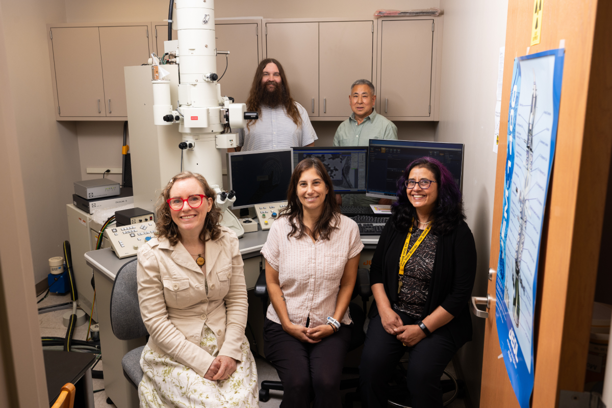
Dr. Andrew Bellemer (back left), Dr. Guichuan Hou (back right), Dr. Sarah Carmichael (front left), Dr. Cara Fiore (middle front), and Dr. Ava Udvadia (front right) have been awarded a two-year, $182,268 grant to upgrade the Transmission Electron Microscope in the William C. and Ruth Ann Dewel Microscopy Facility. Photo by Chase Reynolds
The grant, awarded through NSF's Major Research Instrumentation (MRI) Program, will replace the transmission electron microscope (TEM)'s obsolete camera and update its operating software. As App State's only instrument capable of imaging subcellular structures, microorganisms, nanoparticles, mineral formations, and macromolecules at extremely high resolution, the TEM relies on a functional camera to operate. The new camera will restore full imaging capability, enabling researchers and students to capture high‑speed, high‑resolution images with minimal distortion. Updated software will allow users to stitch images together, analyze samples in three dimensions, and expand the scope of research questions that can be addressed with the instrument.
The TEM supports research and teaching across multiple scientific fields at App State. With restored imaging capabilities, faculty and students will be able to advance investigations in plant development, fruit fly neurobiology, cave sediment composition, mineral characterization, optic nerve regeneration, and muscle recovery following injury. These projects span biology, geology, environmental science, and materials science, reflecting the TEM's role as a multi‑user instrument that supports interdisciplinary research and student training.
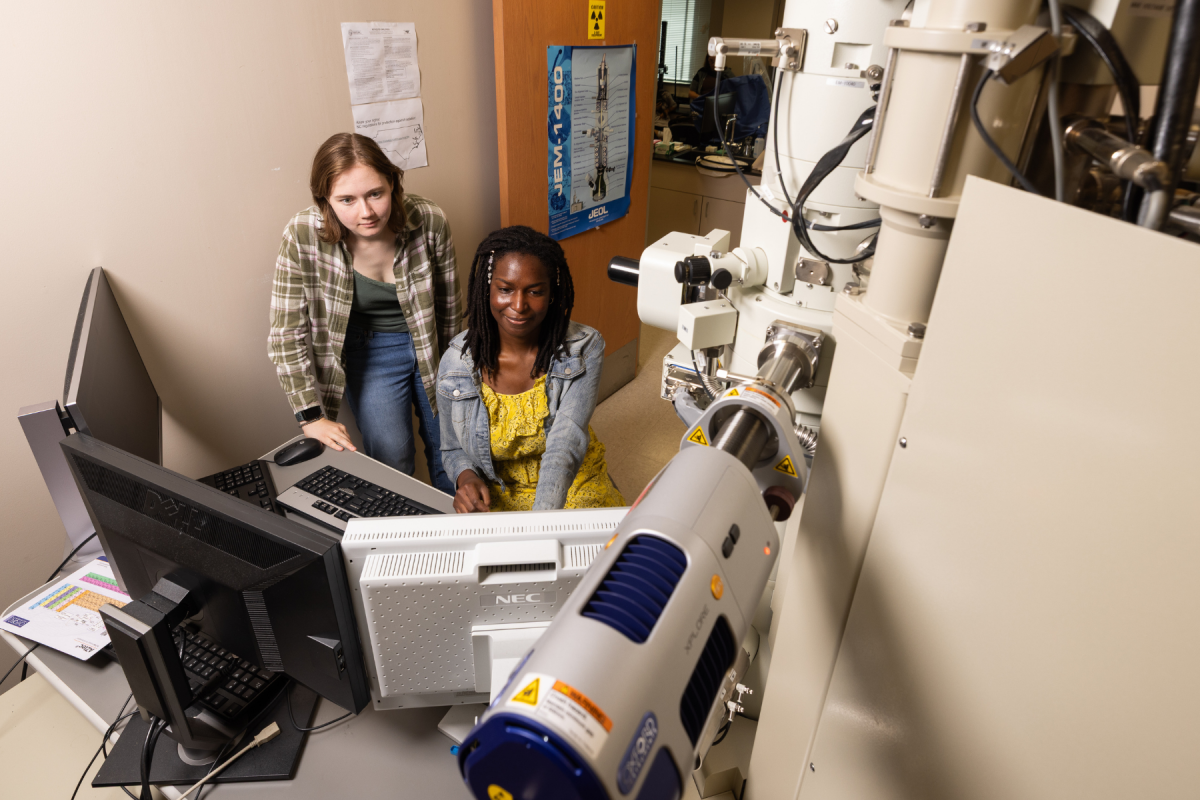
Biology graduate students Kya Hargan (left) and Olivia Carter ’25 (right) learn how to use the Transmission Electron Microscope in the William C. and Ruth Ann Dewel Microscopy Facility. Photo by Chase Reynolds
The TEM was originally acquired through a previous MRI award in 2009, also led by Hou. That instrument — a JEOL JEM‑1400 — has supported more than a decade of research and teaching across biology, physics, geology, chemistry, and materials science. The new camera will extend the microscope's lifespan by at least 10 years.
MRI awards expand access to advanced scientific instrumentation at U.S. universities. At App State, the TEM is central to hands‑on research training, giving students experience with industry‑standard imaging tools. The upgraded camera and operation software will ensure continued access to high‑resolution microscopy for both undergraduate and graduate researchers.
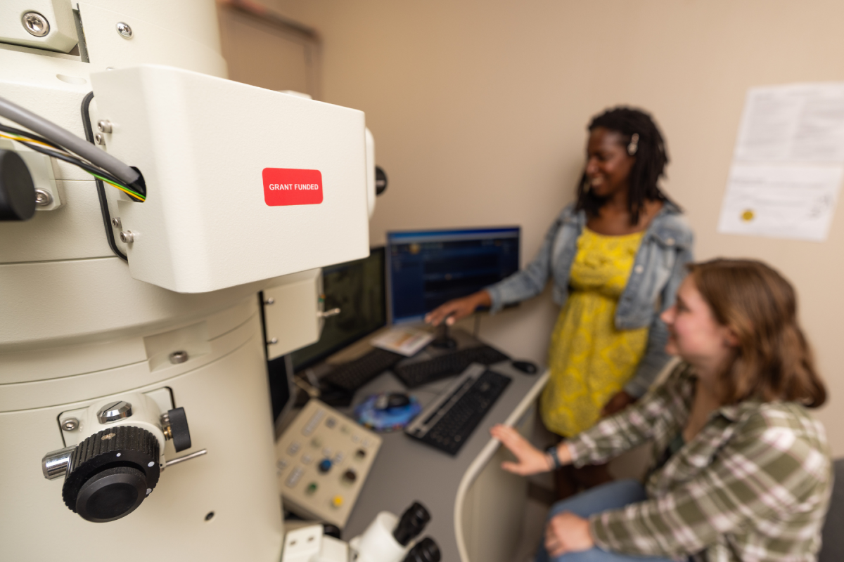
The Transmission Electron Microscope in the William C. and Ruth Ann Dewel Microscopy Facility was acquired through a previous Major Research Instrumentation (MRI) grant in 2009, also led by Dr. Guichuan Hou. Photo by Chase Reynolds
###
About the William C. and Ruth Ann Dewel Microscopy Facility
The William C. and Ruth Ann Dewel Microscopy Facility in Appalachian State University's College of Arts and Sciences provides shared light and electron microscopy resources for research and teaching across campus. Located in the Rankin Science Building, the facility offers imaging services, training, and support for faculty, undergraduate students, and graduate students, and fosters collaboration among departments and regional partners. Learn more about the Microscopy Facility
About the College of Arts and Sciences
The College of Arts and Sciences (CAS) at Appalachian State University is home to 17 academic departments, two centers, and one residential college. These units span the humanities and the social, mathematical, and natural sciences. CAS aims to develop a distinctive identity built upon our university's strengths, traditions, and locations. The college's values lie not only in service to the university and local community, but through inspiring, training, educating, and sustaining the development of its students as global citizens. More than 6,800 student majors are enrolled in the college. As the college is also largely responsible for implementing App State's general education curriculum, it is heavily involved in the education of all students at the university, including those pursuing majors in other colleges. Learn more about the College of Arts and Sciences
By Lauren Gibbs
September 1, 2026
BOONE, N.C.
Five researchers pose in a laboratory beside a large microscope and computer monitors, smiling warmly in a collaborative, welcoming setting.
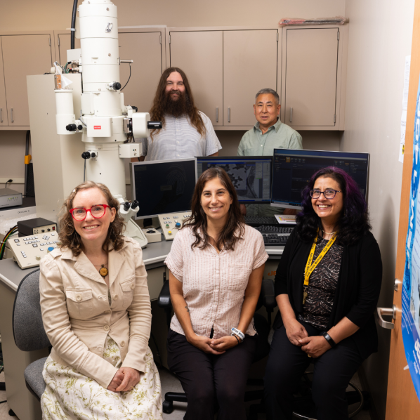
Dr. Andrew Bellemer (back left), Dr. Guichuan Hou (back right), Dr. Sarah Carmichael (front left), Dr. Cara Fiore (middle front), and Dr. Ava Udvadia (front right) have been awarded a two-year, $182,268 grant to upgrade the Transmission Electron Microscope in the William C. and Ruth Ann Dewel Microscopy Facility. Photo by Chase Reynolds